
Protuberance Anatomy
External occipital protuberance Protuberantia occipitalis externa. Definition. The under surface of the jugular process is rough, and gives attachment to the Rectus capitis lateralis muscle and the lateral atlantoöccipital ligament; from this surface an eminence, the paramastoid process,sometimes projects downward, and may be of sufficient.
external occipital protuberance Archives
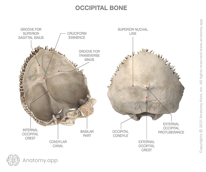
The occipital squama is the larger, more posterior portion of the external occipital bone. It is a thin, flat bone that forms the base of the skull and contributes to the formation of the posterior cranial fossa.The occipital squama is marked by several prominent features, including the external occipital protuberance, the external occipital crest, and the superior nuchal lines.
The Physical Effects of Technology Saratoga Spine
External occipital protuberance (EOP) is a midline bony prominence in the occipital bone that ligamentum nuchae and trapezius muscle attach to its tip that named Inion. The tentorium cerebelli attaches to its internal surface. Entheses are the sites of ligament, tendon or joint capsule attachment to the bone..
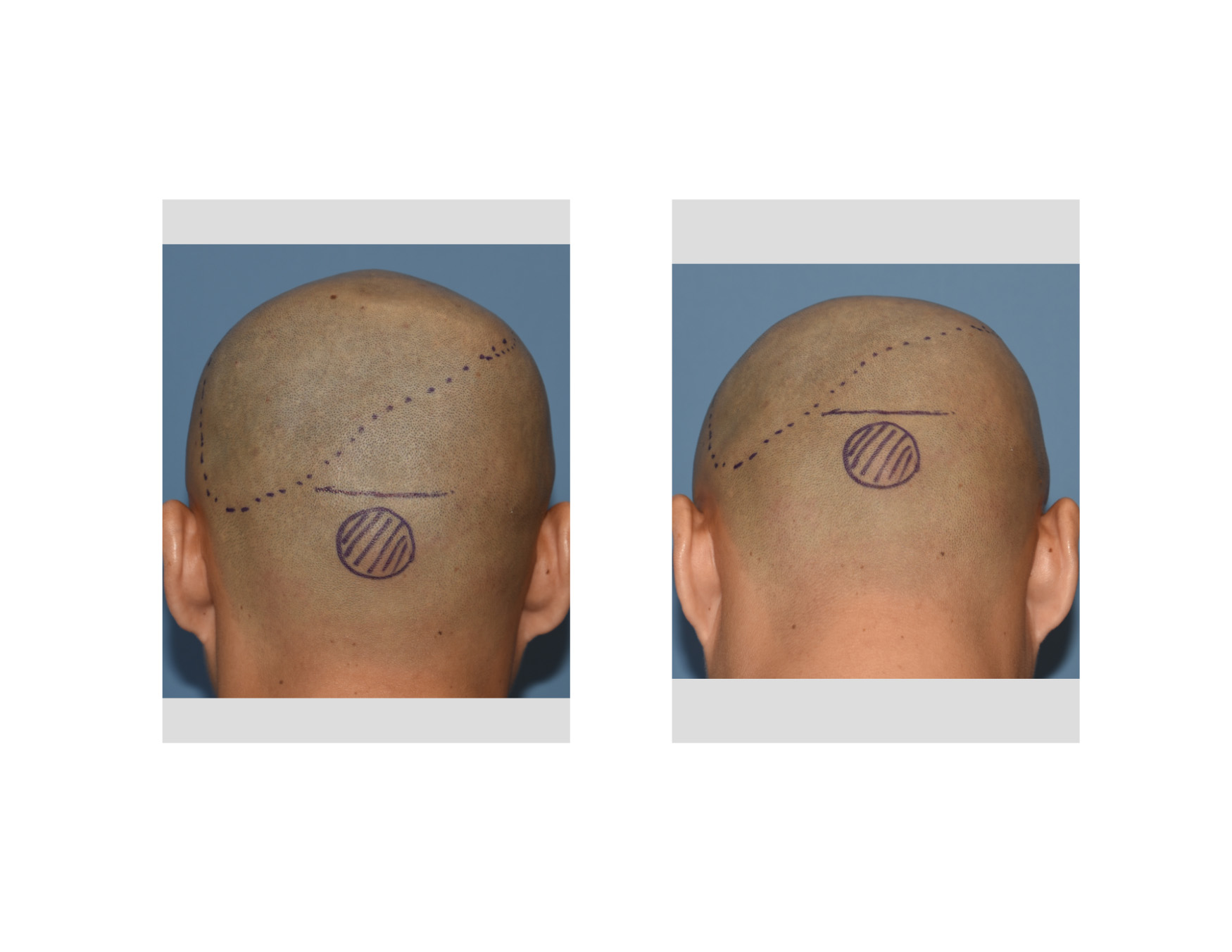
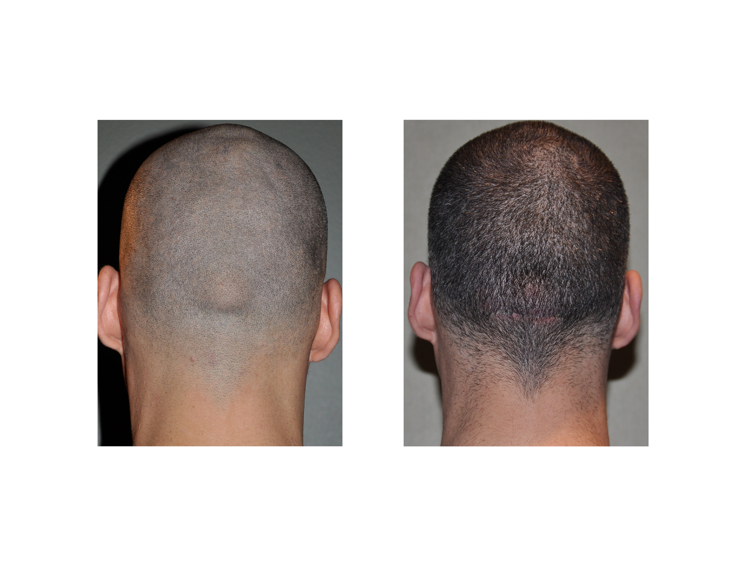
Plastic Surgery Case Study Custom Occipital Implant with Occipital Knob Reduction Explore
External Occipital Protuberance (EOP) is an anatomical structure located on the occipital bone's posterior surface, at the superior nuchal line level. It is the insertion site of the nuchal ligament and the trapezius muscle [6, 16].

Study Young People are Developing ‘Hornlike Spikes’ at Back of Their Skull Due to Poor Posture
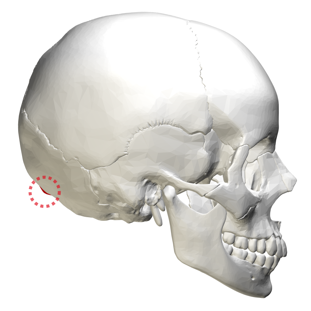
Occipital spurs, also called as occipital knob, occipital bun, chignon or inion hook, is an exaggerated external occipital protuberance (EOP). It is frequently discussed in anthropological literature as a Neanderthal trait but hardly reported and considered as a normal variant in medical literature. It is a frequent finding among males and hence a prominent occipital spur is often used in.
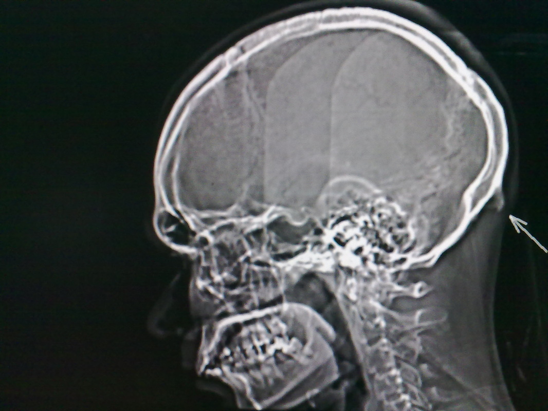
Prominent External Occipital Protuberance Radiology
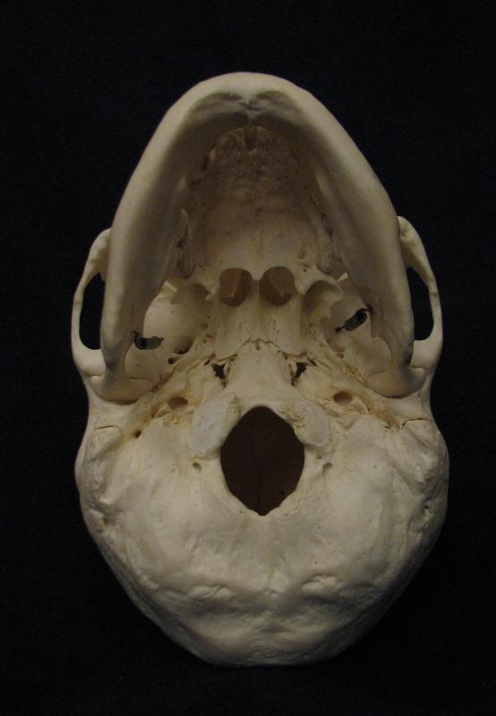
Superiorly, the C1 vertebra articulates (forms a joint) with the occipital condyles of the skull. Inferiorly, C1 articulates with the C2 vertebra, and so on. Below these are the 12 thoracic vertebrae, designated T1-T12. The lower back contains the L1-L5 lumbar vertebrae.. out to the external occipital protuberance. It supports the skull.
VB News Desk Horns?! Nope, just an enlarged external occipital protuberance.
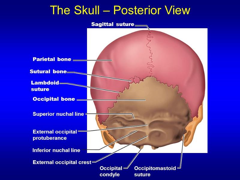
At its midline is a prominence called the external occipital protuberance, with its highest point termed the inion. The superior nuchal line makes an intersection with a vertical midline ridge of bone called the medial nuchal line, which is also known as the external occipital crest, forming an uppercase "T" on the surface of the occipital.
External Occipital Protuberance Inion My XXX Hot Girl
There is a small ridge of bone which arises from the squamous part of the occipital bone known as the external occipital crest. It acts as a site of attachment for the nuchal ligament. The parietal bones are difficult to visualise from the inferior view of the skull, however they can be seen articulating with the temporal and occipital bones.

External occipital protuberance projecting as downward curved horn presenting with intractable
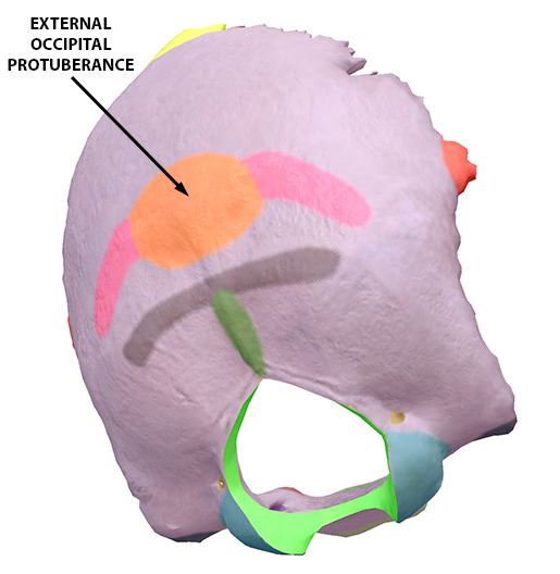
Extends from the midline of the occipital bone towards the lambdoid sutures on either side. Site of attachment of the epicranial aponeurosis. Superior nuchal line: Follows a similar trajectory to the supreme nuchal line but extends further. Marked in the midline by a palpable prominence - known as the external occipital protuberance.

External occipital protuberance (Protuberantia occipitalis externa); Image Yousun Koh Anatomi
Near the middle of the squamous part of occipital bone is the external occipital protuberance, the highest point of which is referred to as the inion. The inion is the most prominent projection of the protuberance which is located at the posterioinferior (rear lower) part of the human skull. The nuchal ligament and trapezius muscle attach to it.
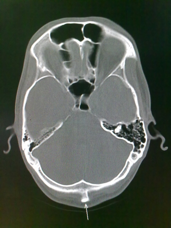
Prominent External Occipital Protuberance Radiology
Anatomy. A midline bump on back of head, on the external surface of satellite-dish-like vertical part (squama) of occipital bone, halfway between the foramen and its highest point. The highest nuchal lines run laterally from the protuberance, with the superior nuchal lines located slightly below.

Physical alignment do you have it? Take a minute & check. — LessWrong
Introduction: In controversial fashion, the presence of an enlarged external occipital protuberance has been recently linked to excessive use of handheld electronic devices.We sought to determine the prevalence of this protuberance in a diverse age group of adults from two separate time periods, before and approximately 10 years after the release of the iPhone, to further characterize this.
Occipital bone Encyclopedia Anatomy.app Learn anatomy 3D models, articles, and quizzes
The bony skull bump — known as an external occipital protuberance — is sometimes so large, you can feel it by pressing your fingers on the base of your skull.
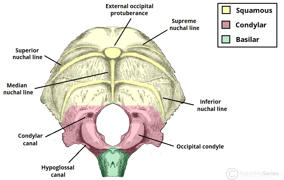
The Occipital Bone Landmarks Attachments TeachMeAnatomy
The external occipital protuberance is the palpable prominence found along the external aspect of the squamous part of occipital bone. It is located at the point along the midline where the occipital and nuchal planes meet. It consists of the inion, which is a craniometric point located at the tip of the external occipital protuberance. The.
What Is the External Occipital Protuberance? (with pictures)
The squamous part is the largest of all four and contains both internal and external surfaces. A palpable prominence known as the external occipital protuberance lies on the midline of the external surface which serves as an attachment for the trapezius muscle.. Furthermore the external surface features three curved lines referred to as nuchal lines:

External Occipital Protuberance Earth's Lab
The external occipital protuberance is a raised bump from the posterior most part of the occipital bone. Extending laterally from it on either side are the superior nuchal lines. The inion is the highest part of that protuberance. Sutures of the skull The lambdoid.
